The original theme of this blog concerned scientific topics geared to college or graduate students. Though I have diverged from this once or twice, this essay returns to the original goal. It involves the intersection of physics and biology and concerns an issue that has periodically occupied my thoughts since I retired.
Introduction: The essay concerns intracellular fluorescent indicators1 which were developed late last century and have proven invaluable tools in cellular physiological research. One person responsible for this breakthrough technique was Roger Tsien 2, who received the Nobel Prize in 2008, along with Osamu Shimomura and Martin Chalfie 3, for the discovery and development of green fluorescent protein.
Dr. Tsien was also a preeminent pioneer in developing other chemical compounds used in fluorescent calcium imaging. He modified EGTA, a selective calcium-binding compound, making it a more rigid structure, BAPTA, which retains its Ca selectivity but is less sensitive to pH. Then to BAPTA he added fluorescent reporter groups. The resulting compounds, fura-2, indo-1, fluo-3, change their absorption and emission spectra upon binding calcium 4. Thousands of studies have used these and similar compounds, and cellular physiology has been greatly advanced by this impactful scientist.
As is always the case for any physiological probe or measurement device, the question arises, “does the probe change the physiology of the cell or the process which is being measured”? In this case, the answer is likely to be no; however, the details are informative and constitute the subject of this essay.
Background: Cell physiology mainly focuses on signaling; for example, a signal for muscle to contract, for a gland to release a hormone into the blood, or for a neuron to release communication molecules (neurotransmitters) onto the surface of adjacent neurons. These signaling processes are essential in any physiological activity.
The signaling agent in all the previously mentioned examples is a rise in intracellular calcium activity. The free intracellular calcium concentration (the calcium activity) is usually exceptionally low, a tenth of a micromolar or lower. But when a signal arrives, calcium activity may spike to a hundred times this value.
Part of the calcium spike comes from an intracellular Ca store 5, a reticular arrangement of calcium storage compartments. There are other roles of the endoplasmic reticulum, but we consider only calcium storage and release. Once calcium has been released, it moves back into its storage compartment. This must happen rapidly to prevent a prolonged Ca signal from damaging the cell.
A substantial portion of the ER-released calcium attaches to mobile intracellular calcium-binding proteins, which are abundant and naturally occurring in all cells6. These substances are also known as natural calcium chelators or buffers.
Questions: 1.) “Do the endogenous calcium-binding proteins have a role in returning calcium to the storage compartments within the ER? and 2.) Do the exogenous mobile calcium chelators, artificially added, disturb natural signaling”? Yes, to the first question; probably no, to the second.
Related to question 2.): Why do exogenous fluorescent calcium chelators do little to disturb normal calcium signaling? Answer: Excess endogenous calcium buffers make the amount of added exogenous fluorescent chelators negligible.
Related to question 1.): Though not having much effect in intact cells because of excess endogenous buffers, how, under experimental conditions in which these endogenous buffers have been stripped away, does added chelator enhancement of calcium transport occur? As it turns out, this unexpected result is independent of the specific calcium chelator that is used. What are the consequences for calcium homeostasis?
Chelators neutralize calcium’s divalent charge, increasing its lipophilicity⁶. This effect is non-specific, that is it occurs with any calcium chelator, regardless of type.
The mechanism of this chelator effect makes use of the unique nature of the approach site on the calcium pump: chelation facilitates the calcium “feed rate” to the pump. Theoretical calculations suggest, however, that Ca buffers should not be expected to facilitate delivery of ionized calcium to a small calcium binding site 7. Nonetheless, when suitable conditions are present, calcium buffers have shown this effect clearly. Removing the endogenous buffers allows calcium chelators to increase calcium delivery to Ca pumps and the ER. 8,9,10,11,12
Millions of years of evolution have rendered calcium pump structures with calcium pump entry sites designed for maximum efficiency.
The three-dimensional calcium pump crystal structure was published by Toyoshima, Nakasako, Nomura, and Ogawa (2000) and Toyoshima and Nomura (2002). It reveals that the Ca entry site is surrounded by hydrophobic regions of the pump structure13,14. The position of the site has unexpected consequences. It lies at the interface of hydrophobic and hydrophilic regions of the transport molecule, which is embedded within the lipid membrane. The free calcium ion is, of course, hydrophilic, preventing it from entering a hydrophobic region. However, being bound, or chelated, makes the portion of intracellular calcium that is mobile and chelated favorably shielded for finding its way to the Ca entry site. The abundant chelated calcium, together with the position and nature of the calcium entry site, enables more efficient calcium delivery back into the ER.
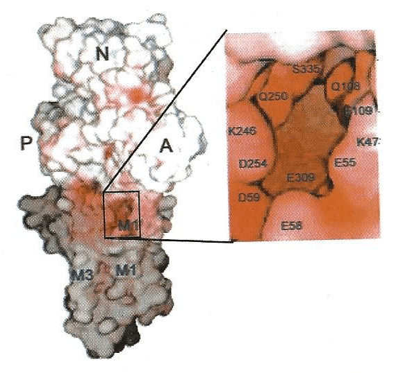
Figure. White represents the hydrophilic surfaces of the pump, exposed to the cell’s aqueous cytoplasm. Grey represents the hydrophobic portions of the surfaces of the pump residing within the lipid of the endoplasmic/sarcoplasmic reticulum membrane. The Ca2+ entry site is shown in the expanded box. The P, M, and A domains (hydrophilic) are generally positively charged. The M1 and M3 represent portions of transmembrane alpha helices. The lettered symbols in the enlarged inset to the right represent identified amino acids and their numbered positions in the protein sequence of the pump molecule. The enlarged inset for the Ca channel inlet shows its structure and position near the Ca binding site, which is deeper within the pump protein. This image is taken from supplementary material of reference (14).
A theoretical analysis of the calcium delivery (or the feed-rate enhancement) by calcium chelators is presented in reference (11).
Conclusion: Calcium chelators boost Ca uptake in microsomes, but not in intact cells, due to the differing experimental conditions. Within living cells, various natural calcium buffers tend to dominate over any artificial ones introduced for imaging. By comparison, when membrane vesicles are isolated, the natural calcium chelators are no longer present. This makes the effects of added chelators easily observable. To better understand cellular physiological processes, intact cells are the appropriate system. To better understand the mechanism of intracellular calcium transport, isolated membranes are more appropriate.
References
1 Sheng Wu, Cheng, Wu, Li . (2024) Comprehensive review of indicators and techniques for optical mapping of intracellular calcium ions. Cereb Cortex. 34(8):346.
2 Roger Y. Tsien – Facts – NobelPrize.org
3 The Nobel Prize in Chemistry 2008 – NobelPrize.org
4 Bailey, Macardle .(2006) A flow cytometric comparison of Indo-1 to fluo-3 and Fura Red excited with low power lasers for detecting Ca(2+) flux. J Immunol Methods. 311(1-2):220-5
5 Blaustein. (1988) Calcium transport and buffering in neurons. Trends in Neurosciences 11(10) 438-43
6 Parsegian. (1969) Energy of an ion crossing low dielectric membrane: solution to four relevant electrostatic problems. Nature 221 844-46
7 Abercrombie, Moore. (2008) Ca Chelators and membrane transport: “Geometric” considerations. The Open Enzyme Inhibition Journal 1_1-4
8 Berman. (1982) Stimulation of calcium transport of sarcoplasmic reticulum vesicles by the calcium complex of ethylene glycol bis (ϐ-aminoethyl ether)0N,N’-tetraacetic acid. J Biol Chem 257(4) 1953-7
9 Timmermans, Bindels, van Os. (1995) Stimulation of plasma membrane Ca2+ pump by calbindin-D28k and calmodulin is additive in EGTA-free solutions. J Nutr 125 (Suppl): 1981S-6S
10 Feher, Fullmer, Wasserman.(1992) Role of facilitated diffusion of calcium by calbindin in intestinal calcium absorption. Am J Physiol 262(Pt1): C517-26
11 Moore, Abercrombie. (1996) Calcium chelators enhance 45Ca accumulation in permeabilized synaptosomes and in microsomes. Am J Physiol 270(2 Pt 1): C628-35
12 Sarkadi, Subert, Gardos. (1979) Effects of calcium EGTA buffers on active calcium transport in inside-out red cell membrane vesicles. Experientia 35(8) 1045-7
13 Toyoshima, Nakasako, Nomura, and Ogawa. (2000) Crystal structure of the calcium pump of sarcoplasmic reticulum at 2.6 A resolution. Nature 405(6787): 647-55
14 Toyoshima and Nomura. (2002) Structural changes in the calcium pump accompanying the dissociation of calcium. Nature 418(6898): 605-11

Leave a comment